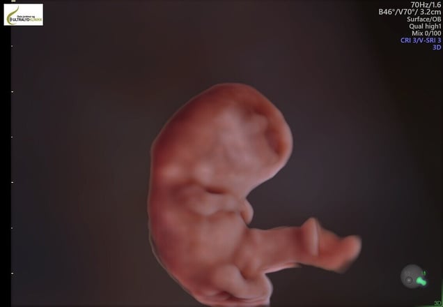
The small embryo begins to move around and measures 23 -32 mm. The weight is a whopping 1 - 2 grams. Both the eyes and ears have been strangely placed on the head, but this week they are moving to where they will be.
The body becomes longer and the head is straightened a little so that the neck becomes clearer.
The arms consist of shoulders, upper arms, elbows, forearms and palms. The legs develop a little more slowly, but towards the end of the week they are divided into thighs, knees, calves, ankles and soles on the feet.
The examination is performed vaginally and takes 30 minutes. We do this examination vaginally in 2D, but if the parents wants us to, we can switch to 3/4D and create fantastic lifelike images.
- Book appointment
- +47 95 41 47 40

Ultrasound weeks 6-7
Before the first 6 completed weeks, the foetus has not developed enough, and we cannot therefore expect to see/hear a heartbeat. Few days play a big role in this context. If you book an appointment before the safe six weeks have passed, the examination might not be able to confirm a heartbeat.
Read more

Ultrasound week 8
This week the brain grows rapidly and the two hemispheres become more distinct. During this week, fingers, toes and elbows begin to form but are difficult to see with a vaginal ultrasound.
Read more

Ultrasound week 11
The overall structure of the brain is now completed. From now on, the brain will further develop specialised structures. However, this does not mean that the brain is fully developed: important substructures in the rough division are not structurally present this early, and it is still a long time before the brain function as it would normally do.
Read more

Ultrasound 12-14
The risk of spontaneous abortion is now greatly reduced. Most miscarriages occur during the first 12 weeks of pregnancy. From now on, this is less common. The foetus's organs are now fully formed, and it is usually serious abnormalities in fetal development that cause the pregnancy to end in miscarriage before week 12.
Read more

We send pictures/videos to your phone
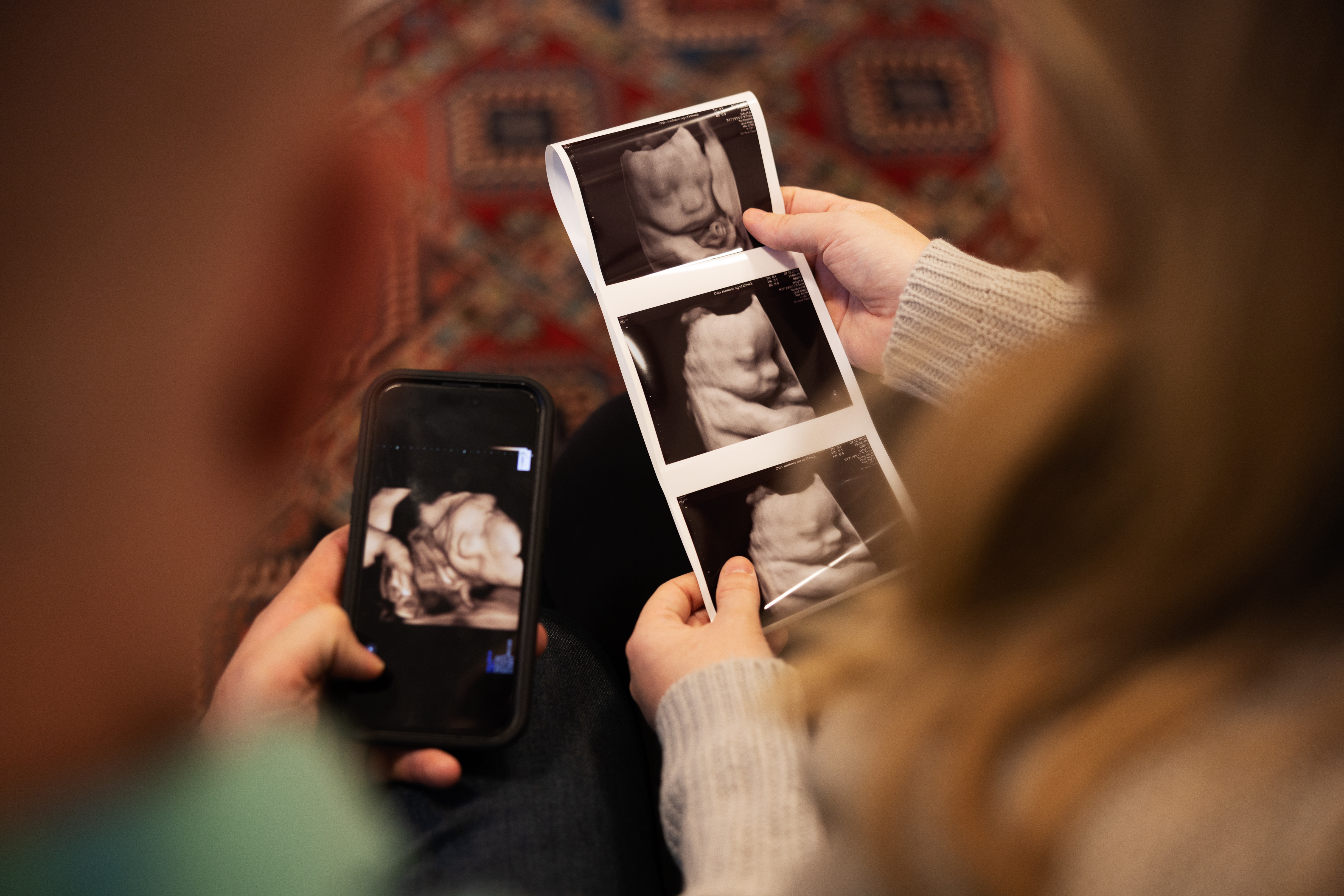
All ultrasound examinations include photos and films sent directly to your mobile phone(s). (Unfortunately, the files do not come with audio)
It’s nice if you bring a companion with you. The experience can be somewhat diminished if there are too many people present. This has to do with both space and potential noise issues. Our experience is that children under the age of 5–6 don’t have the proper understanding or appreciation of the ultrasound examination.
You can easily book an appointment that fits your schedule in our calendar.
FAQ
What is ultrasound?
2D (two-dimensional) ultrasound is sound waves that are transmitted from a probe into the body. The sound waves have such a high frequency that they’re inaudible to the human ear.
When the sound waves hit the body tissue, an echo occurs. The echo causes the sound waves to return to the sound head, which captures these sound signals.
After processing on a computer, the incoming audio signals appear as vivid images in black and white on a screen.
Is ultrasound dangerous?
Ultrasounds of pregnant women have been performed for more than 40 years. No adverse effects on women or foetuses have been recorded.
What happens if a missed abortion (MA) is detected at an early ultrasound?
Sometimes it happens that we do not see cardiac activity. This is usually due to a chromosomal abnormality.
We measure the embryo and refer the woman to the hospital she belongs to. Normally she will get an appointment in a few days. There, a gynaecologist will confirm the finding and a surgical or medical abortion will be performed.
The next day we send her a message to check in on her.
In case of suspicion of illness or abnormalities, what happens next?
The midwives who perform ultrasound have completed one year of foetal diagnosis at the National Centre for Emergency Medicine at NTNU. If we see pictures that deviate from the normal we will refer the woman to a maternal-foetal medicine specialist at the National Hospital. She’ll then get an appointment within a few days.
When is ultrasound done internally (vaginal), and on the stomach (abdominal)?
We perform ultrasound vaginally early in the pregnancy, that is, from weeks 6–12. This is because the embryo is so small and the pictures become much clearer on an internal ultrasound.
The woman must lie with her legs in the leg holder with a towel over her. Most people have no problems with this.
Occasionally we also do an internal ultrasound in week 12. This is if the woman has a uterus that is turned slightly backwards or if she’s overweight.
In the case of a 3D ultrasound, the examination is done abdominally.
.jpg?width=200&height=117&name=OJUK%20logo_m%20bindestrek%20(002).jpg)