.jpg?width=200&height=117&name=OJUK%20logo_m%20bindestrek%20(002).jpg "Oslo jordmor og ultralydklinikk")
A 3D image captures a static moment, while 4D imaging records dynamic movements and videos. Our advanced ultrasound technology enables the creation of vivid, lifelike representations of your child's appearance, movements, and facial expressions.
Equipped with one of the finest ultrasound devices available, our system boasts an HD live function with an exceptional reproduction method.
Conducted when the child is in the optimal position, this examination provides an enriching experience for both parents and the attending midwife.
Recommended between weeks 26-31 (or weeks 24-28 for twins), 3D/4D ultrasound offers the most favorable conditions during this timeframe. However, it is important to note that the technical complexity of the examination may pose challenges, and the assurance of high-quality 3D images cannot be guaranteed.
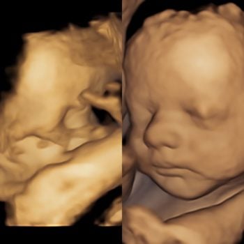
The foundation for successful imaging lies in obtaining a clear 2D image. Adequate amniotic fluid around the face, the child facing upwards towards the mother's stomach, and unobstructed views of the face from arms and legs are essential prerequisites.
The picture shows optimal conditions VS not so optimal conditions.
Our comprehensive examination encompasses the head, brain, spine, neck, heart, stomach, kidneys, bladder, arms, and legs. Additionally, we assess the quantity of amniotic fluid, measure the head, waist, femur, and calculate the child's weight.
Expectations for 3D/4D ultrasound vary among couples seeking this service. While both parents and ultrasound practitioners share excitement at the outset, the quality of the images is contingent upon various conditions, often beyond our control.
Optimal room conditions, characterized by ample amniotic fluid in front of the face, facilitate the imaging process. Conversely, limited space between the fetus and the placenta/womb wall can impede imaging. Fetal position, whether the nose is turned towards the mother's stomach or facing downwards, also significantly influences the outcome.
In instances where conditions are suboptimal, we transition to a 2D examination, performing a thorough well-being check. This includes measurements for growth, amniotic fluid quantity, and umbilical cord blood flow. We meticulously examine fetal organs and provide detailed explanations of the 2D images to parents, ensuring a comprehensive assessment.
If challenges persist, we may ask the mother to reposition or introduce stimuli like juice or cold water to prompt the baby's movement. Despite our relentless efforts, we cannot guarantee favorable 3D/4D imaging outcomes. However, the assurance of the child's well-being remains a paramount consideration, allowing parents to leave the clinic with peace of mind.