.jpg?width=200&height=117&name=OJUK%20logo_m%20bindestrek%20(002).jpg "Oslo jordmor og ultralydklinikk")
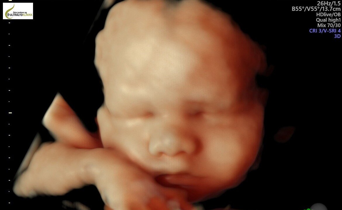
3D and 4D Ultrasound Explained
A 3D ultrasound captures still images, while 4D shows moving images. This advanced type of ultrasound allows us to create lifelike images of your baby, showcasing its appearance, movements, and even facial expressions.
We use one of the most advanced ultrasound machines available on the market, designed to produce high-quality 3D/4D images. The system features an HD live function that provides exceptional image rendering.
When the baby is in an ideal position, this examination becomes a memorable experience for parents and the midwife alike.
Recommended Timing for 3D/4D Ultrasound
We recommend scheduling a 3D/4D ultrasound between weeks 26 and 31 of pregnancy when the conditions inside the womb are most favorable. For twins, we suggest scheduling between weeks 24 and 28.
It’s important to note that despite the baby’s gestational age, this examination can sometimes be technically challenging. Several conditions must align for successful images, and as such, we cannot guarantee high-quality 3D images.
Ideal Conditions for a Successful Examination
The process begins with a clear 2D image. To achieve good results:
- The baby should have enough amniotic fluid surrounding its face.
- The baby should be facing upward, toward the mother’s abdomen, with no arms, legs, or umbilical cord obstructing the view.
During the examination, we also evaluate the baby’s head, brain, spine, neck, heart, stomach, kidneys, bladder, arms, and legs, as well as the amount of amniotic fluid. Measurements of the head, abdomen, and femur are taken to estimate the baby’s weight.
Managing Expectations for 3D/4D Ultrasound
Many expectant parents look forward to 3D/4D ultrasound, eager to see lifelike images of their baby. At our clinic, we share this excitement, but the outcome depends on the conditions at the time of the examination.
Good conditions yield great images, while less ideal conditions can result in less clear pictures. Neither we nor the parents can predict the baby’s position before the examination begins.
What Do We Mean by “Conditions”?
Room inside the womb plays a significant role:
- If there is ample amniotic fluid in front of the baby’s face, capturing clear images is much easier. Conversely, when the space between the baby and the placenta or uterine wall is limited, the imaging becomes more challenging due to the "tight" conditions.
The baby’s position is also important:
- If the baby is facing upward, toward the mother’s abdomen, imaging is more straightforward. If the baby is facing downward, toward the mother’s back, capturing clear images is more difficult.
Obstructions can also impact the quality of images:
- If the baby has a hand, foot, or the umbilical cord in front of its face, it can block the view. For instance, we might see a hand covering one eye or only half the face. If the baby is lying sideways, we may only capture a glimpse of an ear.
Ultimately, the baby decides how the session will go— the “little boss”! 😊

Non-optimal Optimal
If conditions are favorable, we transition quickly from 2D to 3D images and 4D video.
This is incredibly exciting! When we know in advance that the images will turn out well, we share the same joy as the parents over the amazing results. We never become jaded—each session feels special, and we get just as caught up in the moment. We love introducing expectant parents to their much-anticipated baby.
Sometimes, expectations are sky-high, and as ultrasound midwives, we can feel the pressure. Despite our best efforts, there are times when creating clear images or videos of the baby is simply impossible. When conditions are right, it only takes a few seconds to capture stunning pictures. It’s all about finding a little "window" to take the images.
What Do We Do When Conditions Aren’t Optimal?
The image above illustrates optimal vs. non-optimal conditions.
When the baby isn’t ideally positioned—for example, when the nose is facing the mother’s back, pressed against the uterine wall or placenta, or when arms and legs cover the face—it naturally becomes more challenging to create meaningful images.
In such cases, we typically begin the examination in 2D. During this stage, we assess the baby’s position, measure the head, abdomen, and femur to estimate growth, evaluate the amount of amniotic fluid, and check blood flow in the umbilical cord. We also perform a detailed review of the baby’s organs and explain the 2D images to the parents. This part of the process is called a "well-being check." Regardless of whether 3D images are possible, every ultrasound session includes this essential check in 2D.
If conditions aren’t favorable after the well-being check, we may ask the mother to move around to see if the baby shifts position. Drinking juice or cold water can sometimes prompt the baby to turn. Within a few minutes, everything can change, and suddenly the conditions are much more favorable.
Our Commitment to 3D/4D Imaging
We work hard and with great dedication to produce beautiful 3D/4D images, but we cannot guarantee results. Some parents leave the clinic without the images they hoped for, but they leave with the most important reassurance: knowing that their baby is healthy and thriving. 😊

Ultralyd uke 6-7
Ultralyd disse ukene gjøres vaginalt. Vi ser at fosteranlegget ligger på riktig plass i livmoren og at hjertet slår med en takt på rundt 130 -160 slag per minutt. Vi kan se hvor mange fostre kvinnen bærer på og måle fosteranlegget (CRL = Crown-Rump Length), og slik beregne hvor mange uker gravid kvinnen er.
Les mer

Ultralyd uke 9
Det lille embryoet begynner å bevege seg og måler 23 -32 mm. Vekten er hele 1 – 2 gram. Både øynene og ørene har vært litt merkelig plassert på hodet, men denne uken flytter de seg dit de skal.
Kroppen blir lengere og hodet rettes litt opp slik at hals og nakke blir tydeligere.
Les mer
Ultralyd uke 11
Hjernens overordnede struktur er nå på plass. Fra nå av vil hjernen videre utvikle spesialiserte strukturer. Dette betyr likevel ikke at hjernen er ferdig utviklet: viktige substrukturer i den grove inndelingen er ikke strukturelt tilstede så tidlig, og det er fortsatt lenge til hjernen fungerer slik den normalt kommer til å gjøre.
Les mer
Ultralyd uke 12-14
Risikoen for spontant å abortere er nå kraftig redusert. De fleste spontanaborter skjer i løpet av de 12 første ukene av svangerskapet. Fra nå av er dette mindre vanlig. Fosterets organer er nå ferdig dannet, og det er som regel alvorlige feil i fosterutviklingen som gjør at svangerskapet ender i spontanabort før uke 12.
Les mer

Ofte stilte spørsmål
Hva er ultralyd?
2D (2 dimensjonal) ultralyd er lydbølger som sendes inn i kroppen fra et lydhode. Lydbølgene har så høy frekvens at de ikke er hørbare for det menneskelige øret.
Når lydbølgene treffer kroppsvevet, oppstår et ekko. Ekkoet gjør at lydbølgene kommer i retur til lydhodet, som fanger opp disse lydsignalene.
Etter bearbeiding i en datamaskin vises de innkomne lydsignalene som levende svart-hvitt bilder på en skjerm.
3D ultralyd gir tredimensjonalt og dermed et naturtro bilde. Et 3D bilde er et stillbilde og 4D viser bilder i bevegelse.
Er ultralyd farlig?
Ultralyd av gravide har vært utført i mer enn 40 år. Det er ikke registrert skadevirkninger på kvinner eller foster. Det er ingen bivirkninger, ingen data som viser at ultralyd skader fosteret og det er ikke økt abortrisiko ved ultralydundersøkelser.
Man bør likevel tenke på at ultralyd skal utføres av helsepersonell med spesiell opplæring innen ultralyddiagnostikk slik at eksponering holdes så lav som praktisk mulig.
Hvordan fungerer ultralyd?
Ultralydapparatene vi bruker heter Voluson E10, og Voluson E8, maskiner fra General Electric Healthcare som lager svært gode 2, 3 og 4D bilder, og såkalte HDlive visninger. HDlive er en ekstraordinær gjengivelsesmetode som gir realistiske, naturtro bilder av fostre. Gjennom bruk av en avansert belysningsmodell støtter HDlive teknologien skygger, lager en virtuell lyskilde og benytter avansert gjengivelsesteknikk for å vise hud. HDlive teknikken er forskjellig ved at den beregner forplantning av lys gjennom hud og vev. Disse apparatet regnes for å være de beste og mest avanserte som finnes på markedet. Se video av Voluson E10
Hvordan beregne termin?
Lurer du på hvor mange uker gravid du er?
Registrer første dag i siste menstruasjon på denne siden.
Vi benytter avansert teknologi
Ultralydapparatene vi bruker heter Voluson E10, og Voluson E8, maskiner fra General Electric Healthcare som lager svært gode 2, 3 og 4D bilder, og såkalte HDlive visninger. HDlive er en ekstraordinær gjengivelsesmetode som gir realistiske, naturtro bilder av fostre. Gjennom bruk av en avansert belysningsmodell støtter HDlive teknologien skygger, lager en virtuell lyskilde og benytter avansert gjengivelsesteknikk for å vise hud. HDlive teknikken er forskjellig ved at den beregner forplantning av lys gjennom hud og vev. Disse apparatet regnes for å være de beste og mest avanserte som finnes på markedet. Se video av Voluson E10
